defining characteristics
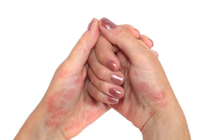
Heliotrope rash (periocular edema + violet color) on sun- exposed areas; Gottron’s papules (red, thickened plaque-like rashes on extensor surfaces), nailbed hemorrhages, violaceous erythema (shawl sign), dilated capillary loops of proximal nail folds, cutaneous calcinosis (ROCK hard)
SYMMETRIC WEAKNESS OF PROXIMAL MUSCLES (usually
lower extremities first, then upper extremities)
disease development
Humoral immune process against vascular endothelium, resulting in the deposition of C5b-9 MAC from complement —
> CD4+ T cell and B cell response –> ischemic muscle injury
DM = DZ of body attacking blood vessels around muscle, causing watershed inflammation around muscle.
potential causes
Complement mediated ischemic muscle injury
epidemiology
1/100,000
F>M
Kids (more calcinosis) & adults
risk factors
labimaging
Histology? Perifascicular atrophy, inflammation of dermal-epidermal jx (interface dermatitis)
Histology of Gottron’s papules? Increased stratum cornum thickening, interface dermatitis
Elevated muscle enzymes
Myositis specific Abs (Anti Jo-1- worse prognosis, Anti Mi-2 -better prognosis)
Bx evidence? Necrosis, upregulation of MAC around blood vessels, regeneration, varied fibers, inflammation around blood vessel (not in the muscle fibers) (CD4+ T/B cells)
XR? Calcinosis
MRI? Muscle inflammation
conventional treatment
corticosteroids
Methotrexate, azathioprine (LT) IV immunoglobulins
photoprotection
complications
Increased risk of malignancy in
+/-4 yrs before/after dx Interstitial lung disease
Diaphragm/ intercostal weakness –> resp arrest
Cardiac rhythm disturbance
prevention
age/gender appropriate cancer screening


