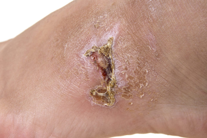
defining characteristics
Xerosis, erythema, red-brown discoloration from hemosiderin deposits and degraded extravasated RBCs, dilated superficial veins; often involves medial ankle, possible lipodermatosclerosis (hard feeling from underlying fat necrosis); hypopigmentation, ulceration
disease development
Abnormal circulation in skin comprises the skin barrier, causing dryness and inflammation –> itch –> LSC & SD
OLD theory – SD caused by stasis and hypoxia, but pts actually have high flow rate and oxygen
Abnormal microcirculation – increased permeability of dermal capillaries allows leakage of fibrinogen, which polymerizes to fibrin to form fibrin cuff around capillaries –> ultimately
inflammation
potential causes
epidemiology
risk factors
Venous insufficiency
labimaging
conventional treatment
Often require hospitalization for tx of venous ulcers
complications
Can be complicated by LSC


